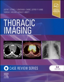
Thoracic Imaging: Case Review Series EPUB
by Justin T. Stowell, Jonathan H. Chung, Jeffrey P Kanne, Theresa C. McLoud, Gerald F. Abbott
Part of the Case Review series
EPUB
Description
Master the critical imaging content you need to know with this thoroughly updated title in the popular Case Review series. Thoracic Imaging offers a highly illustrated, case-based preparation for board review to help residents and recertifying radiologists succeed on exams, demonstrate a clinical understanding of thoracic imaging, and improve imaging accuracy and interpretation. Cases include both common and difficult-to-diagnose disorders spanning the range of diseases impacting the thorax, including sarcoidosis, lymphoma, traumatic aortic injury, small airway disease, idiopathic interstitial pneumonias, developmental lung disease, and lung cancer.
Presents more than 145 high-yield case studies organized by level of difficulty, helping you build your knowledge and confidence in stages. All cases have been refreshed and rewritten to capture the latest clinical implications and diagnostic pearls on thoracic conditions that you will be tested on.
Includes multiple-choice questions, answers, and rationales that mimic the format of certification exams.
Uses short, easily digestible chapters covering the full range of thoracic imaging for efficient, effective learning and exam preparation.
Features hundreds of high-quality, full-color images (many new to this edition) representing a wide range of clinical situations encountered in thoracic imaging. Images include chest radiographs, CT, PET-CT, and MRI to help you expand your visual identification and diagnostic skills.
- Clearly see and interpret 400 high-quality, state-of-the-art images representing a wide range of clinical situations encountered in thoracic imaging.
- Get fresh perspectives from 200 updated or new cases reflecting the most recent changes in thoracic imaging, including PET-CT, emerging pulmonary infections, smoking-related lung diseases, Fleischner guidelines for management of incidentally detected lung nodules, and ground glass and part-solid lung nodules.
- Spend less time searching and more time learning with easy-to-navigate chapters focused on visual identification and diagnosis, and reorganized by degree of case difficulty.
Information
-
Download - Immediately Available
- Format:EPUB
- Pages:352 pages
- Publisher:Elsevier Health Sciences
- Publication Date:29/03/2023
- Category:
- ISBN:9780323449168
Information
-
Download - Immediately Available
- Format:EPUB
- Pages:352 pages
- Publisher:Elsevier Health Sciences
- Publication Date:29/03/2023
- Category:
- ISBN:9780323449168